Radiology View: What’s Your Read?
It can be challenging to interpret dental radiographs in our veterinary patients. Below are radiographs for your evaluation followed by interpretations from MedVet board-certified dentists.
Below are various dental radiographs (figures 1-3). View the x-rays and make your diagnosis.
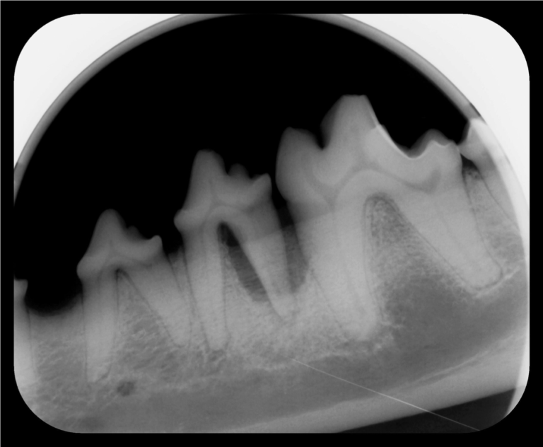
Figure 1. Dental radiograph of the left mandible of a seven-year-old Belgian Malinois. See figure 4 for the interpretation.
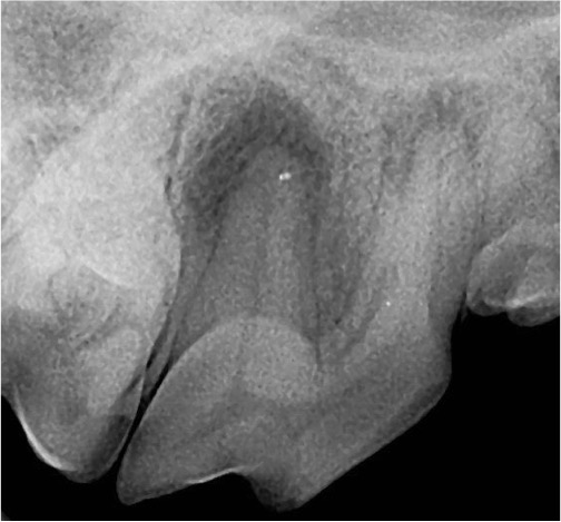
Figure 2. Dental radiograph of the right maxillary fourth premolar of a five-year-old Beagle with an uncomplicated crown fracture. See figure 5 for the interpretation.
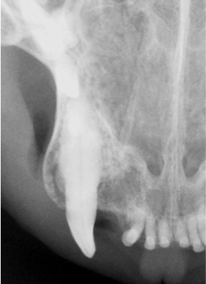
Figure 3. Dental radiograph of the right maxillary canine of a cat with swelling of the gingiva. See figure 6 for the interpretation.
ANSWERS:

Figure 4. Interpretation of figure 1. Vertical bone loss due to periodontal disease of the left mandibular fourth premolar and first molar. The blue line illustrates where the alveolar bone should extend too. The red line illustrates the where the bone has receded due to periodontitis. Vertical bone loss may be treated by bone grafting or extraction of the affected teeth or a combination thereof.
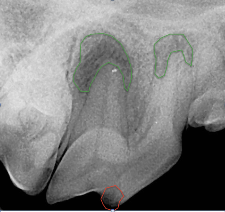
Figure 5. Dog with uncomplicated crown fracture. Interpretation of figure 2. Periapical lucency of the right maxillary fourth premolar secondary to pulp inflammation. The green circles denote periapical lucency which indicate loss of mineral of the alveolar bone secondary to inflammation. The red circle denotes where the crown was fractured. Note, this fracture did not communicate with the pulp yet still led to disease. Treatment options for this tooth are extraction of the tooth or root canal therapy.
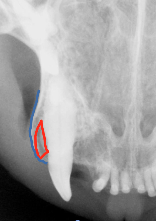
Figure 6. Dental radiograph of the right maxillary canine of a cat with swelling of the gingiva. Interpretation of figure 3. Expansile osteitis of the right maxillary canine. This is a unique pattern of periodontal bone loss seen in cats which grossly appears as a bulbous swelling of the gingiva. The red denotes vertical bone loss and the blue outlines the expanding alveolar bone, which should taper toward the crown when no inflammation is present. Treatment is typically extraction of the tooth. However if there is any concern for neoplasia, tissue should be submitted for histopathology.