Our clients often seek advice on how to treat the messy, frustrating problem of tear staining. If you research “tear staining”, you will find numerous different products, ideas, and suggestions that claim to fix or prevent the staining. The fact that there are so many products available means that there is not ONE magic treatment or approach that will work for every dog.
The red/brown discoloration in tears comes from porphyrin. Porphyrins are iron containing molecules produced when the body breaks down iron. Porphyrins are excreted through the gastrointestinal tract, urine, saliva, and TEARS! All dogs have some porphyrin in their tears, but some dogs have more porphyrin and the staining is always more noticeable in white or light-colored dogs.

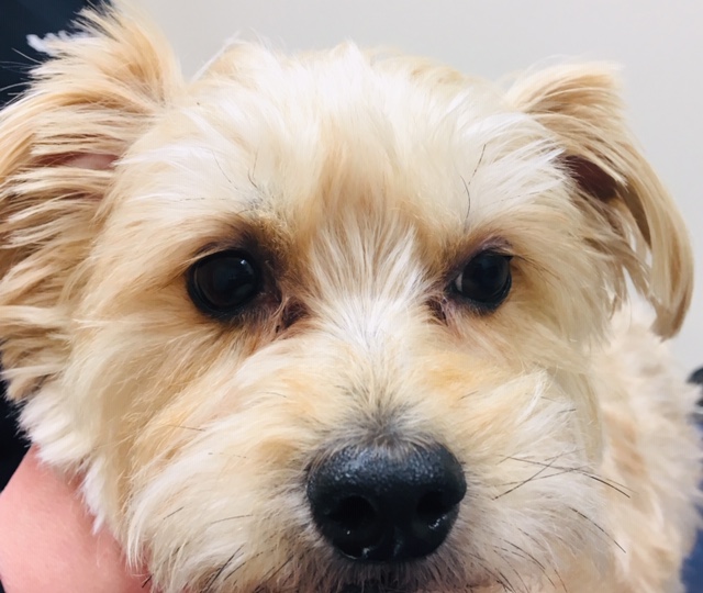
Figure 1. Tear staining in a middle-aged dog.
Causes of Tear Staining
A common misconception about tear staining (Figure 1) is that it is due to excessive tear production. Most dogs with tear staining have normal tear production and do not have an underlying ocular problem. However, many dogs have a normal variation in their eyelid conformation that causes tears to drain onto their face rather than draining down the nasolacrimal puncta and into the nasolacrimal system.
There are three common variations in eyelid conformation that cause tearing onto the face rather than down the nasolacrimal system. They include:
- Tight medial canthal ligament:The medial canthal (palpebral) ligament is tighter in some dogs which causes the medial portion of the eyelids to slightly roll inward. This creates a partial, functionally obstructed nasolacrimal puncta which then causes tears to spill over onto the face.
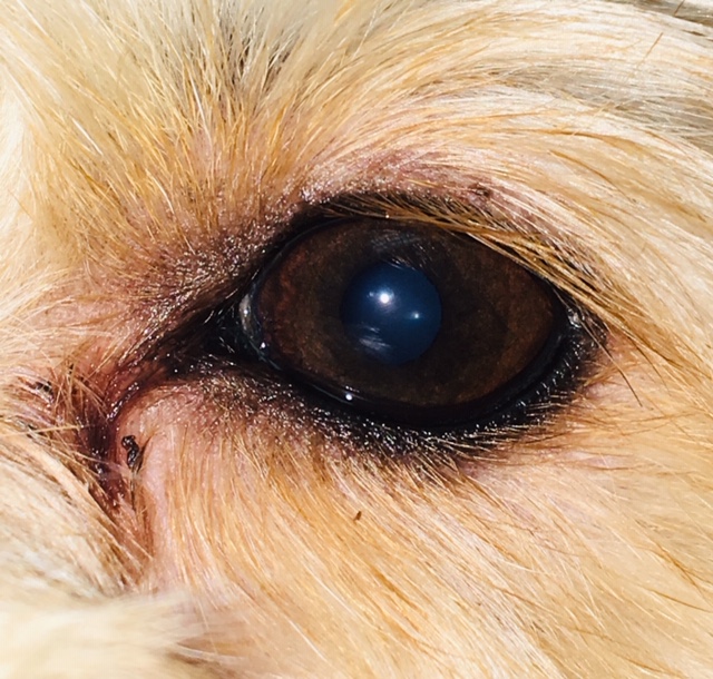
- Haired lacrimal caruncle (Figure 2):The lacrimal caruncle is a triangular prominence in the medial canthus which normally has fine hair and a few sebaceous glands. Some dogs have longer hairs which then wicks tears onto the face bypassing the nasolacrimal puncta.
- Medial canthal troughing (Figure 2):Medial canthal troughing occurs when the skin at the medial canthus forms with a widened trough-like triangular area which funnels tears onto the face, again avoiding the nasolacrimal puncta.

Figure 2. Medial canthal trouging and haired caruncle in a dog.A medial canthoplasty is a surgical option to reduce tear staining in some dogs with these types of eyelid conformations. Cryotherapy can be used to treat a haired lacrimal caruncle. However, in most dogs neither procedure is warranted. Any ocular irritation in a dog with any of these variations in eyelid conformation will cause more than the typical tear staining. Allergies, teething, corneal ulcer or other ocular disease may be the culprit.
Treatment of Tear Staining in Dogs
Some treatment options for dealing with tear staining in dogs include:
- Step one in treating tear staining is keeping the hair around the eyes and nose as short as possible.
- The next step is to keep the face clean and dry. A warm washcloth and baby shampoo are safe to use to clean around the eyes. There are many types of eyelid and eyelash cleaning pads that can also be used to clean the face and around the eyes. Contact lens solution can be used to clean around the eyes—not in the eyes! The boric acid in the contact lens solution oxidizes the iron in the porphyrins and may lighten the staining. After washing the face, always dry the area with a clean towel to prevent ulcerative dermatitis secondary to wet skin.
- Tylosin-containing products claim to treat or prevent tear staining. Tylosin’s effect is unpredictable and often has intermittent efficacy. Because tylosin is an antibiotic, there is controversy using it for cosmesis due to possible drug resistance. There is also controversy regarding tylosin’s addition to over the counter medications that do not always list it as an ingredient or identify how much tylosin is in the product.
- Many probiotic supplements claim to decrease tear staining as well.
Dogs with tear staining should be evaluated to rule out an underlying ophthalmic problem that requires specific treatment. If the tear staining is secondary to conformation, the treatment plan should be an educational discussion focusing on proper cleaning and grooming. Referral to an ophthalmologist is appropriate to rule out underlying ocular disease and to discuss the possible surgical options.

