Veterinary dermatologists primarily care for dogs and cats with conditions involving the skin, ears, hair coat, nails and mucosae. We see parasites, skin infections, allergic diseases, autoimmune diseases, hormone imbalances, and metabolic or systemic diseases that have cutaneous manifestations. Many skin diseases have characteristic features or breed associations we are trained to identify.
The first thing we note at the time of an initial consultation is the signalment (species, age and breed). We inquire about the age of onset of the first problem, what the initial lesions looked like or how it manifested, and how symptoms or lesions have progressed over time. From this information we formulate the list of potential diagnoses which initiates the diagnostic process that leads to a definitive diagnosis. With a diagnosis, the veterinary dermatologist can recommend treatment options.
Why is this historical information important? Some conditions are only identified in certain breeds! One of these is a scaling disorder now recognized in French bulldogs called Zinc responsive dermatosis. Dr. Paul Bloom reported this condition in the breed in 2018 at the North American Veterinary Dermatology Forum (Zinc responsive dermatosis in a French bulldog, Vet Dermatol 2018, 29(4) 267-287). Since that time, I have seen several similar cases.
Variations of Zinc Responsive Dermatosis
There are two variations of Zinc responsive dermatosis in dogs. Type I recognized in Nordic breeds and involves an impairment of zinc absorption from the diet. Type II is recognized in large‐breed puppies and is a deficiency of zinc in the diet or an interaction among components of the diet that results in poor zinc absorption. Lesions of both types may include mild red skin, scaling, crusting, hair loss where there is scale and thickening of the skin. These lesions are found on the ears, elbows, around the eyes, around the lips.
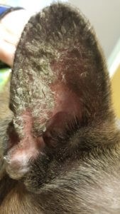
It is speculated that a Type I variation of Zinc responsive dermatosis occurs in the French bulldog. Affected dogs are typically < 3 years of age with lesions characterized by scale forming crusts adhering the concave aspect of the ear pinnae only. Signalment, history, compatible clinical features and biopsy of the skin are used to make the diagnosis. Treatment involves supplementation with elemental zinc. Improvement is recognized in 2 months and resolution is typically appreciated by 3-4 months. Zinc supplementation is continued, perhaps at a reduced dosage, to maintain control of the problem.
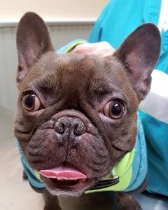
Meet Bubba
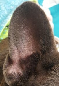
“Bubba” was presented to me at age 3 with a progressive scaling of his ears since age 1. Scaling of the edge of his ears progressed to a crusting on the concave aspect of both ears. Various topical therapies had been tried. He was otherwise healthy primarily indoor dog fed a low fat diet to control a gastrointestinal problem. He was given heartworm preventative and flea and tick preventative monthly year round. The other dog in the household was not affected. After completion of some basic diagnostic tests in the office to rule out other conditions with similar presentation, the diagnosis of Type I Zinc responsive dermatosis of the French bulldog was made. He was treated with zinc methionine orally. In the images provided you can appreciate the lesions before treatment and after four months of zinc methionine supplementation. We will maintain him on zinc methionine once daily.


“Bubba” Type I Zinc responsive dermatosis day 0


“Bubba” Type I Zinc responsive dermatosis day 120 after supplementation with zinc methionine

