Image Courtesy of Wiki Commons
History
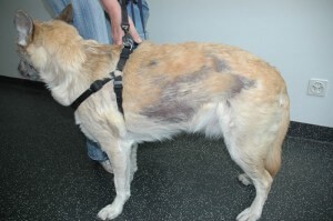
A 10-year-old castrated male Hungarian Vizsla was referred for evaluation of a progressive patchy hair loss that was first noted in November 2012. He had no previous skin problems. There was a mild itch involving the sites of lesions on the trunk and on the forelimbs. The first observation made by the owner was patchy hair loss on the trunk and he appeared to be “moth-eaten.” Scaling of lesions and the entire hair coat followed. The primary care veterinarian performed an evaluation of the alopecic patches with a Woods lamp and this test for dermatophytosis or ringworm was negative. The primary care veterinarian also performed deep skin scrapings to evaluate the pet for demodex mites, and these specimens were negative.
An initial course of antibiotics appropriate for Staphylococcus pseudintermedius, the most common cause of bacterial folliculitis in the dog, did not resolve any of the lesions. A second antibiotic was dispensed and there was no change in appearance of the lesions. An injection of glucocorticoids (steroid) was given followed by an oral glucocorticoid reduced the itch but did not change the appearance of lesions, and lesions were progressing in extent and severity. Upon presentation to the veterinary dermatologist, the dog was on an oral omega-3 fatty acid supplementation. He also has inflammatory bowel disease that is controlled with a controlled diet. The dog was otherwise healthy and up to date on routine vaccinations as recommended by the primary care veterinarian. Of note, no littermates or dogs genetically related to the dog had any history of hair loss.
Dermatologic Examination
Upon initial presentation, there was a “moth-eaten,” patchy alopecia of the entire trunk. The hair at the peripheral aspect of the circular and scaling patches was easily epilated (the hair easily came out). The patches of alopecia on the trunk were characterized by erythema (redness) and with multifocal patches of scale. There were a few dry collarettes and rare 1/2 cm crusts with dry scaling underneath the crust on the head. The limbs, top of trunk, sides of trunk and pinnae (ear flaps) were most affected. The ventral aspect of the body was characterized by a thin hair coat. The dorsal bridge of the nose was alopecic. Oral cavity examination was normal other than mild dental tarter.
Based upon the history and initial examination, the initial differential diagnoses were:
- Superficial bacterial folliculitis
- Adult onset generalized demodex
- Dermatophytosis
- Cutaneous T-cell lymphoma
- Sterile granuloma-pyogranuloma syndrome or other sterile inflammatory skin disease
- Sebaceous adenitis
In- hospital diagnostics performed during the initial consultation:
- Deep skin scrapings were performed to evaluate the dog for demodex. No demodex mites were found. Demodex was ruled out as the cause of the adult-onset patchy hair loss.
- Skin surface cytology specimens were collected and evaluated. There were no bacteria or yeast recovered.
Diagnostic tests submitted to the laboratory during the initial consultation:
- Fungal culture: Skin punch biopsy and hair pluck specimens were submitted to the diagnostic laboratory to evaluate the specimens for the presence of fungal infection, specifically for dermatophytosis. This test was negative and ruled out dermatophytosis as a cause of the adult-onset patchy hair loss.
- Skin punch biopsy specimens were submitted to a veterinary dermatohistopathologist for evaluation of the remaining differentials. The histologic evaluation was consistent with the granulomatous form of idiopathic sebaceous adenitis
Confirmed Diagnosis: Granulomatous idiopathic sebaceous adenitis
Initial Treatment
- Keratolux™ shampoo (Virbac Animal Health): The dog was bathed in this shampoo 1-2 times a week to remove scales.
- Hylyt bath oil spray (DVM Pharmaceuticals): The entire hair coat was misted generously once daily to moisturize the skin surface
- Free form snip tips (DVM Pharmaceuticals): Label dosage based upon body weight was given orally once daily
- Atopica® (Novartis Animal Health): One 100 mg capsule was administered orally every 24 hrs one hour before or two hours after eating.
- Vitamin A Retinol (purchased over the counter)- 8,000 IU given orally every 12 hrs
- Also discussed were once weekly baby oil soaks and the use of a diluted propylene glycol spray 1-2 times daily to effect.
Follow Up
History: The dog was reevaluated three months after the institution of the above treatment plan for a moderately severe case of idiopathic granulomatous sebaceous adenitis. The owner reported that the patchy hair loss lesions on the dog’s trunk had regrown hair and that the scale was fine and minimal on the trunk and limbs. There were no side effects exhibited to any topically or systemically administered medication.
Follow up dermatologic examination: On the dorsal trunk there was a mild fine scaling and there was a slight greasy texture to the slightly thinner than normal hair coat. On the ear pinnae there were a few patches of alopecia and mild scale associated with this patchy hair loss.
Follow-up treatments:
- Atopica® (Novartis Animal Health) 100 mg orally every 24 hrs was continued for 4 additional weeks and then decreased to every 48 hrs. If lesions remain stable, the dosage will be further decreased to every 72 hrs.
- Free Form Snip Tips (DVM Pharmaceuticals) were continued once daily.
- Vitamin A Retinol – 8,000 IU orally every 24 hrs was continued.
- Keratolux™ shampoo (Virbac Animal Health) therapy was continued, but the frequency of bathing was decreased to once weekly.
- Hylyt bath oil spray (DVM Pharmaceuticals) was continued, but the frequency of application of this topical product was decreased to three times a week.
Comments
Sebaceous adenitis is an inflammatory disease that targets the adnexal structures in the hair follicle, specifically the sebaceous gland. It is uncommon in the dog, but has been reported in the Standard poodle, Hungarian vizsla, Akita, Shih Tzu, and Samoyed. In some breeds, an autosomal recessive mode of inheritance is suspected. The heritability pattern in the Hungarian Vizsla is not known. We do not recommend that affected dogs, or those genetically related to affected dogs, be used for breeding purposes.
Most cases of sebaceous adenitis present to the veterinarian for scaling involving the back, neck, head, face, and tail. Lesions may remain localized, become multifocal, or progress to generalized lesions. In short coated dogs, the clinical appearance of lesions is often a patchy to diffuse hair loss, as in this case. It is important to note that this lesion appearance is also very characteristic of other, more common, skin conditions. Therefore, it is important to rule out other causes of patchy hair loss through a battery of simple diagnostic tests.
In this case, deep skin scrapings ruled out a follicular mite (demodex) and cytology ruled out bacterial folliculitis. A fungal culture ruled out dermatophytosis. The best diagnostic test for this condition is dermatohistopathology. In this case, skin biopsy specimens submitted to a veterinary dermatohistopathologist were diagnostic for idiopathic granulomatous sebaceous adenitis. From the results, the veterinary dermatologist was able to institute the treatment plan suitable for the severity of the case and the owner’s ability to adhere to the therapy plan.
Treatment of mild sebaceous adenitis may include daily oral essential fatty acid supplementation in addition to topical therapies with keratolytic and/or keratoplastic properties to control scale. Such topical therapies include shampoos, emollient rinses, and humectant sprays. For more severe cases, 50-75% propylene glycol and water sprays may be formulated and added to the regimen. The author also recommends a regimen of once weekly soaks in diluted baby oil, followed by a degreasing shampoo. Frequency of such baby oil soaks is tapered over time based upon response to the treatment. Systemic therapy is aimed at preventing further destruction of sebaceous glands and maybe restoring some sebaceous gland function. If the biopsy report identifies that significant inflammation targeting the sebaceous glands remains, the veterinary dermatologist may prescribe naturally occurring vitamin A (retinol) and/or modified cyclosporine A (Atopica®, Novartis Animal Health). Additional medications that have been used with varying results include Tetracylcine and niacinamide in combination, prednisone, and isotretinoin or acitretin. The prognosis for dogs with sebaceous adenitis is variable and depends on disease severity. It is not curable, but clinically, lesions can improve and the dog can have an excellent quality of life.