Radiology View: What’s Your Read?
Patient Presentation
A 13-week-old female Labrador Retriever mix presented to the cardiology service of MedVet Akron for evaluation. The owner stated that the puppy “hangs out with and acts like the old dogs” and describe her as being lethargic and coughing sporadically for the past week.
The following radiographs were made.
View the radiographs below and consider the following questions:
- What are your radiographic findings?
- What is your diagnosis?

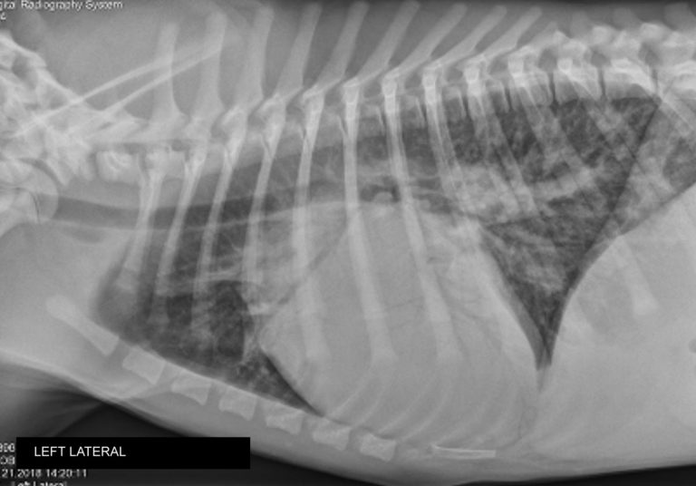
Figure 1. Left lateral thoracic radiograph.

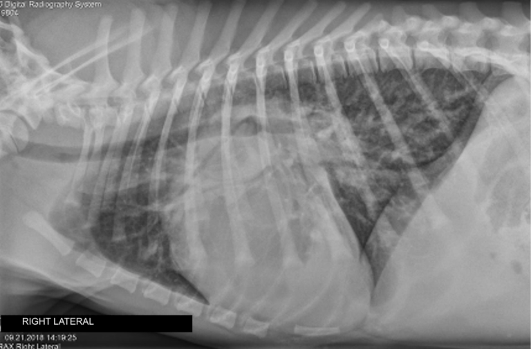
Figure 2. Right lateral thoracic radiograph.

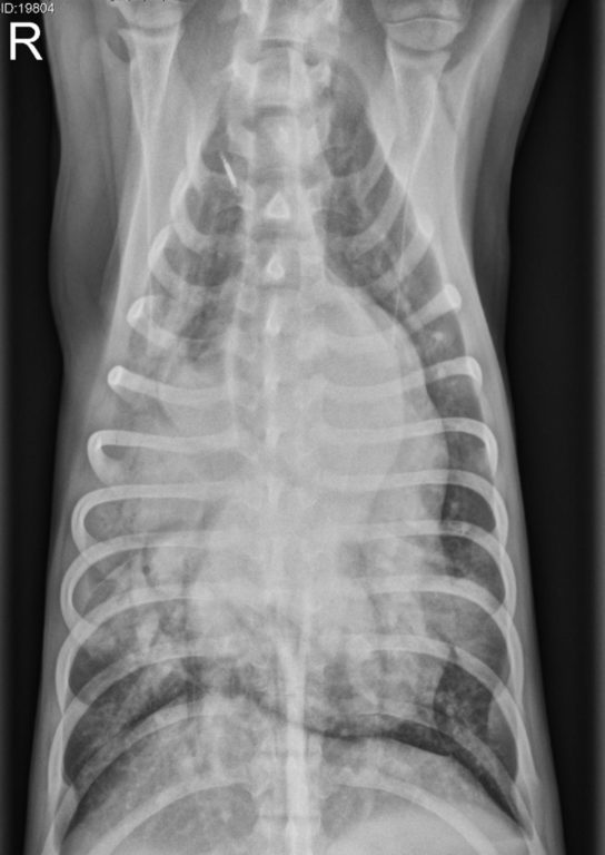
Figure 3. Ventrodorsal thoracic radiograph.
Radiographic Findings
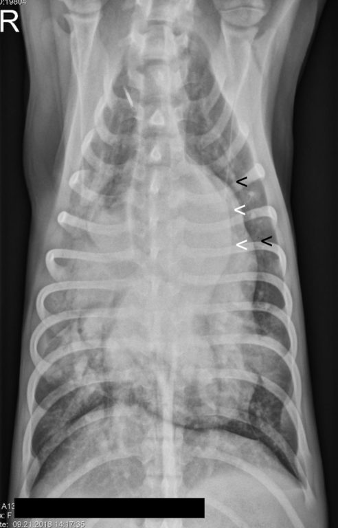
The radiographs show generalized enlargement of the cardiac silhouette but with predominant enlargement of the left cardiac chambers. In the ventrodorsal projection (figure 3) there is enlargement of the aortic arch with bulging at the area of the ductus (white arrowheads) and of the main pulmonary artery segment (black arrowheads figure 4). In the lateral projections (figure 1 and figure 2) the prominent appearance of the heartbase and elevation of the trachea is attributed to enlargement of the aorta and main pulmonary artery segment. The pulmonary vessels are increased in size (more evident in the ventrodorsal projection) and number (more evident in the lateral projections).
There is subtle increase in interstitial opacity of the caudodorsal lung field.
There is alveolar opacification of the right cranial and right middle lung lobes. Note the prominent air-bronchgrams overlying the cardiac silhouette in the left lateral projection. A focal area of alveolar infiltration is also present in the caudal subsegment of the left cranial lung lobe (opacity overlying the cardiac apex in the right lateral projection).

Figure 4. Ventrodorsal thoracic radiograph. Black arrows indicate main pulmonary artery segment.
Discussion
Left-sided cardiac enlargement, enlargement of the aorta (“ductus bulge”), enlargement of the main pulmonary artery segment, and the presence of a vascular pattern (increase in size and number of pulmonary vessels) are all radiographic findings of patent ductus arteriosus (PDA). Echocardiography confirmed the diagnosis.
The subtle increase in opacity of the caudodorsal lung field is suspected to be evidence of early/mild cardiogenic pulmonary edema.
Based on the ventral distribution as well as the severity of the pulmonary infiltration the changes present in the right cranial, right middle and left cranial lung lobes are suspected to be the result of concurrent bronchopneumonia. Although possible it is unlikely that this is a variant of cardiogenic edema.

