A 5-month-old female intact French Bulldog puppy residing in Kentucky presented to her primary veterinarian for lethargy, lymphadenopathy, and fever. The puppy had been recently purchased from a pet store.
A CBC, serum chemistries, and urinalysis were run and the puppy was found to be moderately anemic. The anemia was non-regenerative at that time. The remaining CBC was WNL. Review the blood slides (figure 1 and figure 2) below and consider your interpretation.
What Is Your Interpretation of the Cytology?
The following blood smears were stained with Wright’s (figures 1 – 2) and was directed to a clinical pathologist for review. What is your interpretation?
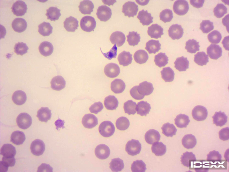
Figure 1. Blood film from a 5- month- old puppy with lethargy, lymphadenopathy, and fever. Magnification 1000X.
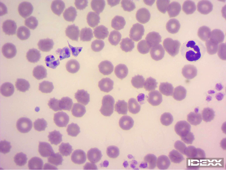
Figure 2. Blood film from a 5- month- old puppy with lethargy, lymphadenopathy, and fever. Magnification 1000x.
The blood film was examined and extracellular spindle shaped structures approximately 15um in size were found. They had a central basophilic staining nucleus and a kinetoplast with an undulating membrane. The structures were compatible with the tryptomastigote form of Trypanosoma cruzi.
What’s Your Diagnosis?
This organism identified above is the etiological agent of Chagas disease which can infect dogs, people, several other wild and domestic mammalian species.
The puppy returned about a week later for re-evaluation. The CBC was repeated and the anemia had improved to a mild anemia and was regenerative. Organisms were still detected upon blood film exam. A lymph node was also aspirated at that time and organisms were identified in the aspirate as well as in the blood. (Follow up information beyond this point was not available.)
CONCLUSION: Chagas Disease in Dogs
Although not common in the United States (US) and primarily seen in Mexico, Latin America, and South America, Chagas infections have been identified in several southern US states, (including Kentucky) with most recognized in Texas.
Transmission requires the appropriate vector infected with the organism and then successful infection of the dog. The vector is a Triatominae, specifically the ‘Kissing bug’. It transmits the organism in its feces which can be ingested by the host, however ingestion of the insect itself is also an effective route of transmission. Other means to infection can occur via blood transfusion, trans-placental, or trans-mammary.
The disease has three stages:
- The acute phase can have a variable manifestation. Some dogs are asymptomatic and others show clinical disease similar to this patient. Parasitemia is usually only evident in the acute phase (the first 3-4 weeks).
- The intermediate (latent) stage is characterized by a positive serology but absence of clinical disease. This stage of disease can last the patient’s entire life, but a percentage of patients (20-30% in humans) may later progress to clinically evident, symptomatic chronic disease (this can occur at any time including years after infection).
- Interestingly, dogs that acquire the disease trans-placentally or trans-mammary more often exhibit clinical disease and have higher mortality rates and shorter survival times in chronic disease stage.
Diagnostics for Chagas Disease in Dogs
Currently, IFA testing is recommend for diagnosis of this disease. Identification of the organism in blood is also diagnostic.
Humans can contract Chagas’ disease but are not as susceptible as dogs to the development of infection. There is no direct transmission from the dog to human, but if a dog is positive, then the people sharing a close environment may have the potential for infection. This disease in people has been described as one of the most lethal endemic infectious diseases in the Western Hemisphere.
Recent studies of dogs tested for disease surveillance reveals approximately 18% of shelter dogs infected and between 7-18% of infection rate of US working dogs (border patrol and military).
An antiparasitic medication (Benznidazole) in the acute phase has shown efficacy, it appears to be less effective in the chronic stage.
In the state of Texas, the Texas Department of State Health Services, Zoonosis Control is requesting notification of infection in dogs.
References
Curtis-Robles, R., Snowden, K. F., Dominguez, B., Dinges, L., Rodgers, S., Mays, G., & Hamer, S. A. (2017). Epidemiology and Molecular Typing of Trypanosoma cruzi in Naturally-Infected Hound Dogs and Associated Triatomine Vectors in Texas, USA. PLoS Neglected Tropical Diseases. https://doi.org/10.1371/journal.pntd.0005298
Machado, F. S., Dutra, W. O., Esper, L., Gollob, K. J., Teixeira, M. M., Factor, S. M., … Garg, N. J. (2012). Current understanding of immunity to Trypanosoma cruzi infection and pathogenesis of Chagas disease. Seminars in Immunopathology. https://doi.org/10.1007/s00281-012-0351-7
Meyers, A. C., Meinders, M., & Hamer, S. A. (2017). Widespread Trypanosoma cruzi infection in government working dogs along the Texas-Mexico border: Discordant serology, parasite genotyping and associated vectors. PLoS Neglected Tropical Diseases. https://doi.org/10.1371/journal.pntd.0005819
Teixeira, A. R. L., Hecht, M. M., Guimaro, M. C., Sousa, A. O., & Nitz, N. (2011). Pathogenesis of chagas’ disease: Parasite persistence and autoimmunity. Clinical Microbiology Reviews. https://doi.org/10.1128/CMR.00063-10
Travi, B. L. (2019). Considering Dogs as Complementary Targets of Chagas Disease Control. Vector-Borne and Zoonotic Diseases. https://doi.org/10.1089/vbz.2018.2325
Greene (2012) Infectious Disease of the Dog and Cat, 4thedition, Snowden and Kjos, p722 -749. St. Louis, MO: Elsevier.
Hondo, C., Rodriguez, J., Curtis-Robles, R., et al. (2019)Repeated cross-sectional study of Trypanosoma cruzi in shelter dogs in Texas, in the context of Dirofilaria immitis and tick-borne pathogen prevalence. Journal Veterinary Internal Medicine.