Hyperadrenocorticism in dogs may occur spontaneously or may be iatrogenic from the administration of glucocorticoids (GC) like prednisone (a steroid). Spontaneous hyperadrenocorticism is pituitary dependent (most commonly a pituitary gland adenoma producing ACTH hormone that signals the adrenal gland to produce cortisol hormone without feedback regulation) or adrenal dependent from an adrenal tumor (tumor produces excess cortisol without feedback regulation). The excess cortisol is responsible for the clinical signs and abnormalities seen with hyperadrenocorticism (“Cushing’s Syndrome”).
Diagnosis
Iatrogenic hyperadrenocorticism is what veterinary dermatologists see most in the face of GC use for the diseases we manage- allergies (canine atopic dermatitis), autoimmune diseases (pemphigus foliaceus), and other immune mediated skin conditions. The diagnosis of iatrogenic hyperadrenocorticism is made based upon history, exam findings, general laboratory work findings (complete blood count, serum biochemistry test, urinalysis, urine cortisol to creatinine ratio), and tests to evaluate the pituitary adrenocortical axis (ACTH stimulation test, low-dose dexamethasone suppression test). Abdominal ultrasounds may also be done as a screening test.
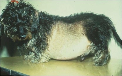
Iatrogenic hyperadrenocorticism is often seen in toy and small breed dogs, but large breed dogs may also be affected. The average age is 10-14 yrs and the average time on a GC medication is 9.4 months. We see this with oral, injectable, and topical use of GC. It may be seen with both appropriate and excessive use of these medications. Signs may include an enlarged liver upon palpation of the abdomen, a “pot belly” appearance of the abdomen, obesity, muscular atrophy, lethargy, excessive water consumption, excessive urinations, and excessive appetite. Skin changes include thinning of the hair coat and skin, prominent cutaneous vasculature, hair loss that may or may not be symmetrical, hyperpigmentation of skin, scaling, bruising, poor wound healing, and calcinosis cutis (mineralization of the skin).
Secondary infections of the skin may occur with bacteria, yeast and some fungi (dermatophyte) due to impaired immune defenses. A mange, demodex mites, may also be a secondary problem. Laboratory work may reveal elevated liver enzymes, cholesterol, triglycerides, and blood glucose. The urine is usually dilute and there may be a urinary tract infection. Complete blood counts also have characteristic features. A definitive test may be done, ACTH stimulation test, and would be consistent with exogenous use of GC by showing a suppressed cortisol concentration at baseline and poor stimulation of cortisol production 1 hour after ACTH administration.
Management of dogs with iatrogenic hyperadrenocorticism involves monitored discontinuation of exogenous GC, treatment of any secondary infections, and monitoring of the laboratory work. Identification and control of the primary disease for which GC were being administered warrants careful review and alternative therapies.
Related Reading: