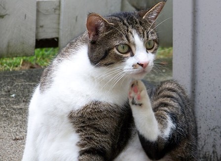
Eosinophilic plaques, granulomas, and ulcers are lesion types that make up the entity of Feline Eosinophilic Granuloma complex. This group of inflammatory lesions, usually associated with an underlying hypersensitivity to fleas (flea allergy), food components (food allergy), or environmental allergens (feline atopic dermatitis) is common in cats.
- Eosinophilic plaques appear as one or few red, raised, eroded plaques. The lesion usually itches and the cat is often observed licking the area excessively. Plaques may occur anywhere on the body, but are usually found on the ventral abdomen, chest and axillae.
- Eosinophilic granulomas appear as coalescing raised swollen, firm, linear plaques that may be papular to nodular, red, and eroded. Granulomas are not typically itchy. They occur anywhere on the body, but are most often found on the inner thigh (linear granuloma), chin or lip. Granulomas can also occur inside the mouth on the tongue or hard palate.
- Eosinophilic ulcers (indolent ulcer, rodent ulcer) appear as small ulcers with raised outer edges on the upper lip near the canine tooth. The ulcer may enlarge and distort the upper lip. They are not itchy.
Diagnosis
To diagnose this problem the veterinarian will consider the history and findings on dermatologic examination to develop a differential list. Diagnostics that may be recommended by the veterinarian are cytology to look for the characteristic cell called an eosinophil and other evidence of infection, skin scrapings to look for demodex mites, and skin biopsies to confirm the diagnosis and rule out other causes of similar lesions (like some tumors/cancer).
Treatment
Secondary bacterial infections that are identified should be treated with appropriate systemic antibiotics for two to four weeks. Treating such an infection may decrease the severity of the lesions by 50% or more. Antihistamines and oral essential fatty acids may be prescribed. Modified cyclosporine (Feline Atopica®, Novarits), or systemic glucocorticoids (prednisolone, dexamethasone or triamcinolone) are additional anti-inflammatory medications used to get lesions in to remission. Glucocorticoids are often used initially after infection is resolved and Atopica® or antihistamines may be prescribed to decrease the likelihood of recurrence whilst the underlying cause of the lesions is pursued.
Cause
Underlying hypersensitivities should always be considered, identified, and controlled if present. First, and often concurrent with treatment of the active lesion, a strict flea control program for all pets in the home to control for flea allergy is instituted. Second, a diagnostic food trial using a diet recommended by the veterinarian is instituted. If flea allergy and food allergy are ruled out as the underlying cause, feline atopic dermatitis should be considered the primary allergy causing lesions. Sometimes, eosinophilic granuloma complex is idiopathic in nature (underlying cause not determined).
Prognosis
Prognosis is variable, depending on the underlying cause. If an underlying hypersensitivity is identified and controlled, successful treatment and control is more likely. If an underlying cause is not determined, cats will require lifelong treatment and should be followed by the veterinarian or a veterinary dermatologist.
Related Reading:

