Canine Atopic Dermatitis (Environmental Allergy) in Dogs
Dogs with canine atopic dermatitis persistently lick their paws, bite limbs, or rub their bodies to itch. Learn how to help your pet.
It’s not unusual for dogs to occasionally lick certain body sites or chew on a leg or paw. But if they are persistently licking their paws, biting their limbs, shaking their heads, chewing their rump, or rubbing their face and ears on things to scratch an itch, such that it affects their activities of daily living, the pruritus (the term for itch) may be a clinical sign of canine atopic dermatitis (CAD).
About Canine Atopic Dermatitis
Canine atopic dermatitis is the genetic predisposition to develop allergic antibodies, meaning they become sensitized to environmental allergens such as pollen, environmental mites, environmental molds, danders, etc. This happens over time as the allergen is absorbed through the skin (percutaneous absorption) and presented to cells of the skin immune system. Allergen absorption through the skin is due to a genetic abnormality in a structural component of the skin. A cascade of events occurs here, and the body produces molecules that drive itch, resulting in itching and inflammation. With re-exposure of the immune system to an offending allergen(s), clinical signs develop.
Canine atopic dermatitis is more prevalent in Terriers, Retrievers, German Shepherds, Bulldogs, Pugs, Lhasa Apsos, Shih Tzus, Boxers, Dalmatians, Spaniels (especially Cocker Spaniels), and Shar Peis.
Signs of Atopy in Dogs
The most common sign of atopic dermatitis is the pruritus, or itch. Secondary infections can also occur. Typical allergic reactions in humans like coughing, sneezing, and runny eyes are rare to absent in dogs. Common allergens include pollen (weed, tree, grass), environmental mites (house dust mites, storage mites), mold spores, or insects.
The inflammation at the skin surface alters the skin’s normal microenvironment. Resident bacterial flora and yeast may overgrow in these favorable conditions and exacerbate itch.
Diagnosing Atopy in Dogs
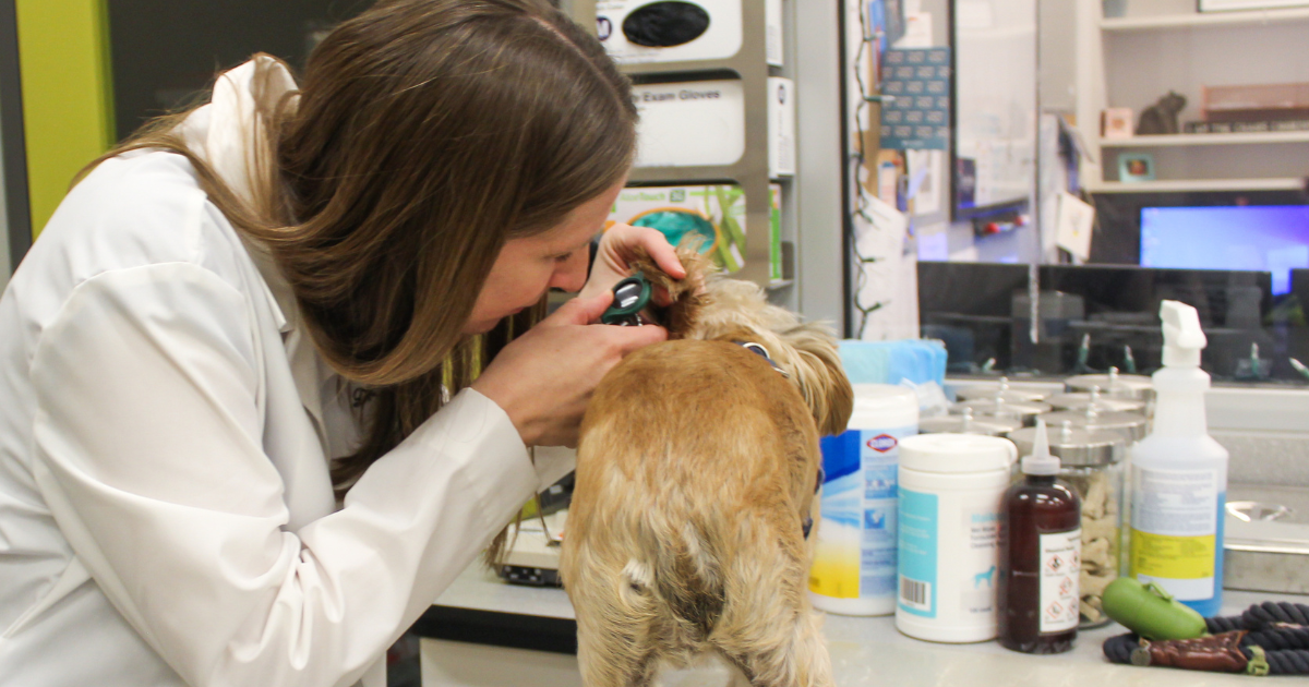
Diagnosing atopic dermatitis is typically straight forward. Your veterinarian will make a clinical diagnosis by ruling out other causes of pruritus/itch in the dog. Any ectoparasites or infections must be treated and controlled. If itch is non-season (year-round) cutaneous, your veterinarian will also explore if there is an adverse food reaction. After considering these different diagnoses, if itching continues of the face, paws, ears, ventrum, limbs, or perineum, a diagnosis of atopic dermatitis can be made.
Historical information about your pet’s condition helps refine the diagnosis. This includes items like the age of onset, course of disease over time, seasonality of signs, type of clinical signs, affected body sites, and response or lack of response to certain medications.
Treatment of Atopy in Dogs
Canine atopic dermatitis treatment is multi-modal, meaning a combination of treatments is typically needed. Treatment is lifelong, as atopic dermatitis is not a curable condition.
Treatment of Atopic Dermatitis
Allergen specific immunotherapy is the treatment for atopic dermatitis. It is the process of administering small doses of allergen to the pet either by sublingual delivery or subcutaneous injection. In time, the immune system becomes tolerant or conditioned to the offending allergen so that, upon re-exposure, the allergic inflammation is reduced.
To determine what allergens to include in the allergy vaccine for your pet, a veterinary dermatologist will perform allergy testing. This may be an intradermal testing (IDT) or serum allergy test. Ideally, both are done. Intradermal testing is performed by injecting small amounts of diluted allergen just under the pet’s skin. If the pet is sensitized to a specific allergen, the injection site will erupt with a “hive” after 5-15 minutes. This then fades over 15-30 minutes.
Pet owners give allergy shots or sublingual immunotherapy drops at home after learning the skill. Response to therapy takes 9-12 months. If there is a good reduction in the observed signs, immunotherapy is continued for life and adjusted over time in consultation with the veterinarian managing your pet’s allergy.
Symptomatic Therapy
Symptomatic therapy is needed to manage clinical signs and help keep your pet comfortable. Options that are recommended may be for acute flares, seasonal signs, or part of the multi-modal long-term treatment plan.
There are many options to consider. Antihistamines, supplements such as high doses of omega 3 fatty acids, glucocorticoids, Apoquel, Atopica, and Cytopoint are all options to mitigate itch and manage skin inflammation. Topical therapy is critically important in taking care of a dog with atopic dermatitis.
Your family veterinarian may recommend that your pet see a veterinary dermatologist who specializes in skin conditions. We partner with your family veterinarian to develop the best treatment plans to improve your pet’s quality of life.
FAQs
Share

Contents