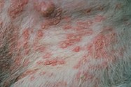
Deep bacterial pyoderma is a bacterial infection involving the deep layers of the skin; the dermis and sometimes the subcutaneous tissue. It usually starts as a superficial infection of the hair follicle (folliculitis) that breaks through the follicular wall into the dermis. The resulting inflammation and infection of the dermis is called furunculosis. When the subcutaneous tissue becomes involved, it is called a panniculitis.
Factors that predispose animals to developing deep bacterial pyoderma include parasitic infestations such as Demodex, hormonal diseases such as hypothyroidism and hyperadrenocorticism, the use of immunosuppressive medications such as steroids, and allergic diseases such as food allergy, flea allergy, and atopic dermatitis.
Diagnosing Pyoderma in Dogs
Clinical signs of deep pyoderma include swelling, discoloration, crusting and ulceration of the skin, large blood-filled swellings called hemorrhagic bullae, and blood-tinged to purulent draining tracts. Lesions may be painful and/or itchy. Lymph nodes may be enlarged and the pet may be anorexic, febrile and lethargic.
Recommended tests initially include cytologic examination of exudates obtained from lesions and bacterial culture and susceptibility testing to identify the bacteria causing lesions. This latter test identifies the appropriate antibiotic to eradicate the infection. Additional testing may be done to try and identify an underlying reason for the infection.
Treatment of Pyoderma in Dogs
Treatment is usually based upon the results of bacterial culture and susceptibility testing and may involve 8 to 16 weeks of an oral antibiotic and topical therapeutics. The involved areas may heal with scarring and may be more prone to reinfection. The prophylactic use of topical therapeutics and addressing the underlying reason for the infection is helpful in preventing recurrence of deep bacterial pyoderma.
Image Source: Vetbook.org
Related reading:

